A lateral tension model for cranial mouse neural tube closure
A lateral tension model for cranial mouse neural tube closure
De La O, J.; Galea, G. L.; Martin, A.
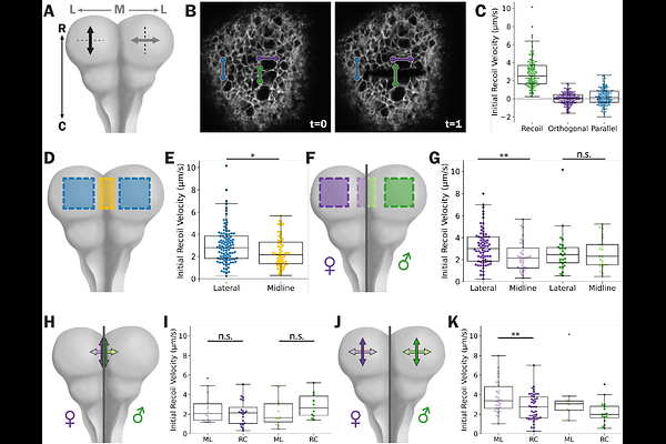
AbstractOrganisms mold their tissues into increasingly more complicated shapes during development using a set of deformation motifs. One set of motifs are tissue hinges and creases which are created through cell-level shape changes. Hinges and creases are often made from active, anisotropic cell constriction at the hinge, which can help drive tissue folding. This contractile hinge model is observed in a variety of developmental tissue folding contexts, like during neural tube closure (NTC) of commonly studied species. However, patterns of cells constriction are inconsistent with this model, in the mouse brain. Additionally, formation of the midline neural tube hinge and crease are insensitive to the molecular machinery needed to induce cell shape changes. Here, we test if a contractile hinge is the driving mechanism for mouse cranial NTC. Through targeted laser ablations, we infer tissue tension in the folding mouse cranial neural tube. In contrast to predications of the contractile hinge model, we find the midline hinge has relatively low and isotropic tension, the lateral neural folds have higher anisotropic tension. We also show that regional patterns of tension vary by sex. We propose a lateral tension model for mouse cranial NTC and theorize on the connection between tissue mechanics and sex in NTC defects.