GFP-Free Live-Neuron Quantitative Imaging Reveals Compartmentalization and Growth Dynamics of PolyQ Aggregates
GFP-Free Live-Neuron Quantitative Imaging Reveals Compartmentalization and Growth Dynamics of PolyQ Aggregates
Bi, X.; Lin, L.-E.; Miao, K.; Wei, L.
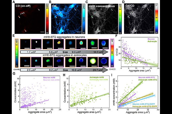
AbstractHuntington\'s Disease (HD), the most prevalent polyglutamine (polyQ) neurodegenerative disorder, features brain aggregates induced by mutant huntingtin (mHtt) proteins harboring expanded polyQ tracts. Despite extensive efforts, molecular mechanisms of polyQ aggregates remain elusive. Here we establish quantitative stimulated Raman scattering imaging of polyQ aggregates (q-aggSRS) for non-invasive investigations in live neuronal co-cultures using deuterated glutamine labeling. Q-aggSRS allows for specific visualization by targeting the distinct Raman peak from carbon-deuterium bonds, eliminating the need for bulky GFP tagging. Coupled with analysis from aggregate-tailored expansion microscopy, newly designed two-color imaging, and pulse-chase visualization, we comprehensively quantified the mHtt and non-mHtt proteins within the same aggregates across varying sizes, cell types, mHtt constructs, and subcellular locations. Our findings demonstrate a two-phase aggregate growth model with a distinct core-shell spatial organization, reveal significant heterogeneity in nucleus/cytoplasm compartmentalization specific to neurons, and identify previously unrecognized \"gel-like\" aggregates specifically in neuronal nuclei. These insights should advance our understanding of native polyQ aggregates and our proposed interaction coefficients may offer new quantitative parameters for developing effective HD therapies.