Fluorescence Characterization of Extracellular Vesicles using Single-Molecule Confocal Microscopy
Fluorescence Characterization of Extracellular Vesicles using Single-Molecule Confocal Microscopy
Zhao, T.; Pelegrina-Hidalgo, N.; Edwards, D. C.; Bak, K. M.; Karmakar, U.; Fernando, A. J.; Vendrell, M.; Rossi, A. G.; Kunath, T.; Cockroft, S. L.; Saleeb, R. S.; Horrocks, M. H.
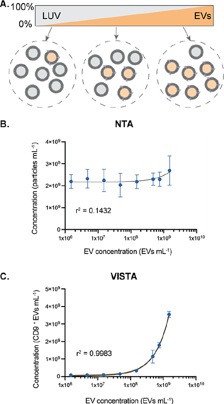
AbstractExtracellular vesicles (EVs) are small, membrane-bound particles released by cells into the extracellular environment. They play a pivotal role in cell communication and have recently gained prominence as biomarkers. However, their low abundance and high heterogeneity challenges their accurate characterization using conventional approaches. To enable the specific detection of individual EVs, we coupled EV-specific antibodies labeled with two different fluorophores with fast-flow microfluidics and single-molecule confocal microscopy. This allowed us to determine the concentration of EVs down to femtomolar levels (~10,000,000 EVs/mL), and we demonstrated the approach\'s capacity to detect EVs even in the presence of other lipid vesicles. We highlighted the ability to quantify EVs in serum and plasma samples, without the need for purification. Furthermore, we compared the yield of EVs extracted from both serum and plasma using ultracentrifugation and various size exclusion chromatography approaches. Overall, our method offers a highly specific, sensitive and easy-to-use solution for characterizing EVs from different sources.