Pericytes in heart
Pericytes in heart
ZHAO, G.; Lederer, W. J.
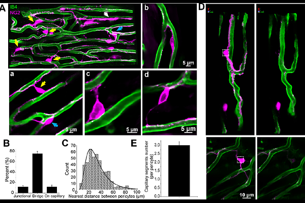
AbstractPericytes are cells associated primarily with capillaries and are thought to play an important role in the regulation of blood flow. They are often referred to as \"mural\" cells because they are so frequently found on the exterior walls of small vessels - particularly the capillaries. In heart, high-resolution real-time observations and measurements of pericyte function under physiological conditions are challenging to obtain because of vascular motion, tissue depth and vigorous functional movement. For these reasons, the heart may be one of the most difficult tissues in which to examine pericyte function. Recently, we introduced a perfused papillary muscle preparation (the Z-Prep) that allows us to observe coronary arteries, arterioles, venules, capillaries and myocytes in real time at physiological temperature and pressure while also imaging pericytes 1, 2. Here we present an initial study intended to visualize and characterize quantitatively cardiac pericytes in heart at physiological pressure and temperature conditions. Vascular anatomy was imaged using a z-stack protocol with a rapidly spinning disk confocal microscope. Here the anatomical organization of the pericytes is shown at high resolution with respect to the microcirculation components and cardiac myocytes. The surprising findings include the high abundance of pericytes in native tissue, the extent of their spread on the capillaries themselves, and the existence of major pericyte extensions that travel intimately along the surface of neighboring ventricular myocytes and attach to capillaries on the distant side. These extensions arise from a capillary-based pericyte location and normally end on another capillary endothelial surface and we have named them \"bridging\" pericytes. Taken together this anatomical organization suggests that the pericytes provide signaling, communication and contractile services to important cellular components of the heart. There is also a provocative suggestion that pericytes in heart are unusually fragile since they suffer an extremely high degree of loss during cellular isolation procedures. However, our investigation of the organization argues against this fragility because of the durability of the dynamic pericyte organization and function despite the stress and brutality of the contracting heart. The work presented here lays the foundation for critical functional studies of pericytes in heart in both health and disease.