Computational aberration-corrected volumetric imaging of single retinal cells in the living eye
Computational aberration-corrected volumetric imaging of single retinal cells in the living eye
Feng, G.; Godinez, D. R.; Li, Z.; Nolen, S.; Cho, H.; Kimball, E.; Duh, E. J.; Johnson, T. V.; Yi, J.
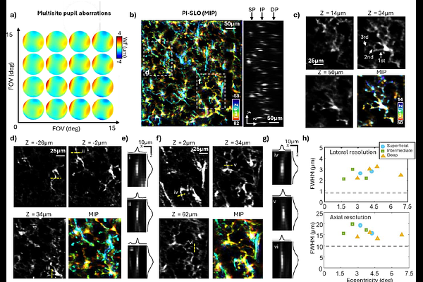
AbstractThe eye offers a unique non-invasive window for accessing single-cell level structures and functions of the central nervous system (CNS) throughout the retina. However, strong and space-varying ocular aberrations, along with limited volume rates, challenge large-scale cellular imaging in living eyes and stymie the full potential of possible biological and pathological studies in retina. Here, we present plenoptic illuminated scanning laser ophthalmoscopy (PI-SLO), a 3D fluorescent retinal imaging modality that enables high-speed, widefield, volumetric single-cell imaging with low phototoxicity. By capturing multiple angular images of fluorescence signals from the entire volume, PI-SLO enables digital aberration correction and 3D imaging across a >20deg FOV with >23 Hz volume rate. We leverage this structural and functional imaging modality to investigate three key aspects of CNS physiology through the living mouse retina, including: microglial process dynamics, vascular perfusion, and light evoked calcium fluxes in inner retinal neurons. PI-SLO is a versatile non-invasive platform for in vivo investigation of retinal and CNS physiology at the cellular level.