Lactate cannot replace glucose for maintaining the viability of mouse and human glioma cells
Lactate cannot replace glucose for maintaining the viability of mouse and human glioma cells
Miller, E. Y.; Duraj, T.; Ta, N. L.; Lee, D. C.; Mukherjee, P.; Shaver, O.; Seyfried, T. N.
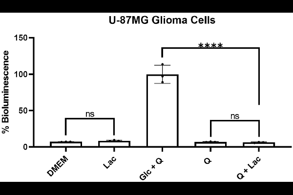
AbstractObjectives: Aerobic lactic acid fermentation (the \'\'Warburg effect\'\') is associated with OxPhos insufficiency and altered energy metabolism in most cancers. Whether lactate is a major fuel in cancer cells remains debated. This study investigated whether lactate could serve as a metabolic fuel in glioma cells and replace glucose to support viability. Methods: A bioluminescence ATP assay and calcein-AM/EthD-III double-staining were used to measure ATP content and viability in mouse (VM-M3, CT-2A) and human (U-87MG) glioma cells differing in cell biology and genetic background. Viability was assessed in thioglycollate-elicited peritoneal macrophages (TPMs) from VM/Dk and C57BL/6J mice, used as syngeneic non-neoplastic controls for VM-M3 and CT-2A gliomas, respectively. Oxygen consumption rate (OCR) was determined using the Resipher system. Results: Lactate alone failed to sustain ATP content and viability in all glioma cell lines. ATP content and viability were lower in cancer cells cultured in glutamine and lactate than in glucose and glutamine. In contrast, lactate alone sustained over 45% viability in both TPM models. Moreover, viability was similar between TPMs cultured in glutamine and lactate and in glucose and glutamine. In human U-87MG, lactate addition under severe glucose restriction increased OCR and viability. A low dose of the glycolysis inhibitor 2-deoxy-D-glucose abolished both increases. Conclusions: Our results suggest non-neoplastic mouse TPMs utilize lactate more effectively than mouse glioma cells. In U-87MG, lactate utilization appears glycolysis-dependent, given its sensitivity to 2-deoxy-D-glucose. In conclusion, our data does not support lactate as a major oxidative fuel for viability in mouse and human glioma cells.