An immunocompetent osteoblastic model of mammary cancer bone metastasis established by syngeneic intratibial injection of PyMT mammary carcinoma cells in FVB/N mice

An immunocompetent osteoblastic model of mammary cancer bone metastasis established by syngeneic intratibial injection of PyMT mammary carcinoma cells in FVB/N mice
Flatt, C. L.; Nano, S. L.; Goyal, R.; Waltz, S. E.; Niebur, G. L.; Littlepage, L. E.
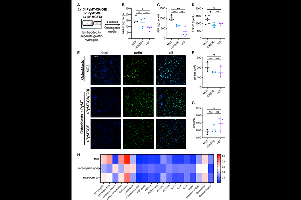
AbstractOsteoblastic bone metastasis, in which disseminated tumor cells drive net bone formation, is a clinically distinct and mechanistically underexplored form of skeletal disease that is enriched in hormone receptor-positive breast cancers. Preclinical models of bone metastasis from breast cancer predominantly rely on immunodeficient hosts inoculated with osteolytic human breast cancer cell lines, limiting the study of immune-dependent mechanisms of bone remodeling. Here we describe the development and characterization of an immunocompetent, syngeneic osteoblastic bone metastasis model using intratibial injection of PyMT-CK(OB), a luciferase-expressing derivative of the MMTV-PyMT mammary carcinoma cell line, in FVB/N mice. PyMT-CK(OB) cells produced detectable bioluminescent signal after intratibial injection, enabling longitudinal monitoring of tumor progression. Micro-computed tomography (microCT) revealed significant increases in trabecular bone volume fraction and trabecular number at three and four weeks post-injection, consistent with osteoblastic remodeling. Histological analysis confirmed dense bone lesion formation in tumor-bearing bones. Critically, this osteoblastic phenotype was entirely absent in immunodeficient NOD SCID hosts, despite robust tumor growth, supporting a role for immune competence in tumor-induced bone formation. Loss of bioluminescent signal in immunocompetent mice reflected either immune pressure on reporter gene expression or limited space for cancer cell expansion in the bone, rather than tumor regression or hypoxia, as confirmed by hypoxia imaging and histological endpoint analysis. In contrast, a second PyMT cell subline, PyMT-CF, maintained sustained bioluminescent signal and produced predominantly osteolytic lesions, providing a complementary syngeneic model of osteolytic disease from the same parental background. In vitro hydrogel coculture experiments and protein array analysis of conditioned media revealed that the PyMT sublines have differing impact on MC3T3 osteoblast mineralization, identifying candidate mediators of divergent bone remodeling phenotypes. R7 mammary carcinoma cells derived from MMTV-RON transgenic mouse mammary tumors did not induce measurable bone remodeling under equivalent experimental conditions. Together, these models provide a validated, immunologically intact framework for studying the mechanistic basis of osteoblastic bone metastasis and evaluating therapeutic interventions targeting the tumor-bone microenvironment.